Boerhaave Syndrome and MalloryWeiss Syndrome (MWS) Lecturio
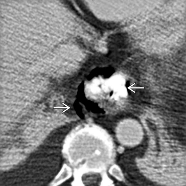
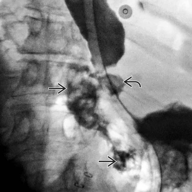
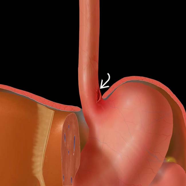
General Features. • Best diagnostic clue. Extraluminal gas and contrast material in lower mediastinum surrounding esophagus. • Other general features. Sudden increase in intraluminal pressure leads to full-thickness esophageal perforation. Left side of distal thoracic esophagus. - Most vulnerable (due to lack of supporting mediastinal.

Cureus Atypical Presentation of Boerhaave Syndrome With Hypoxia and
Boerhaave syndrome is a spontaneous longitudinal perforation of the esophagus due to forceful emesis first described by Hermann Boerhaave in the 18th century. This pathology is best treated with definitive repair and mediastinal and/or pleural drainage procedures. 2 articles feature images from this case 27 public playlists include this case

Boerhaave syndrome Radiology Case
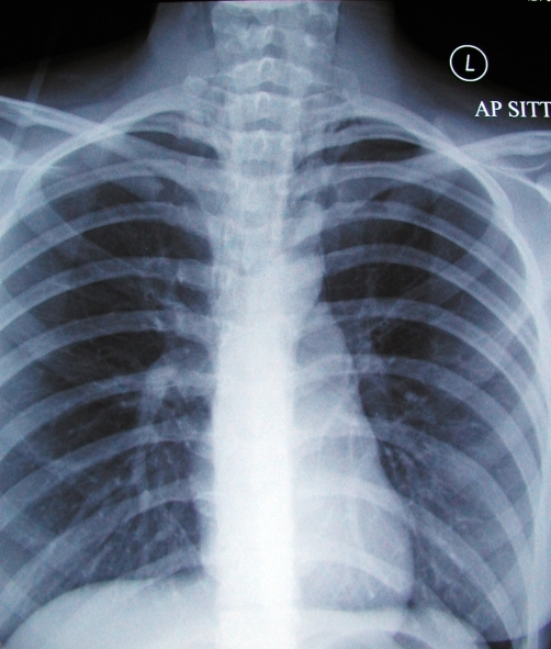
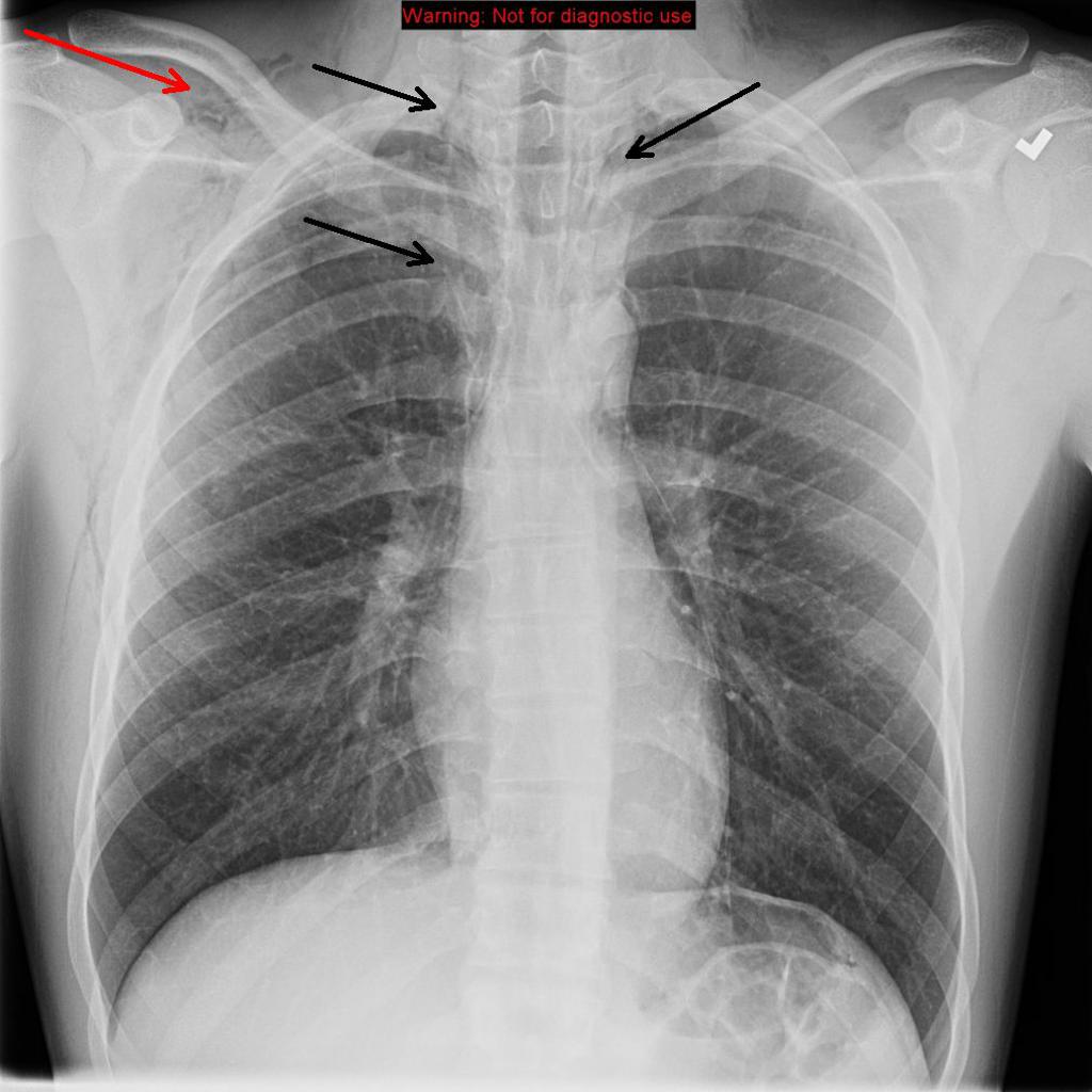
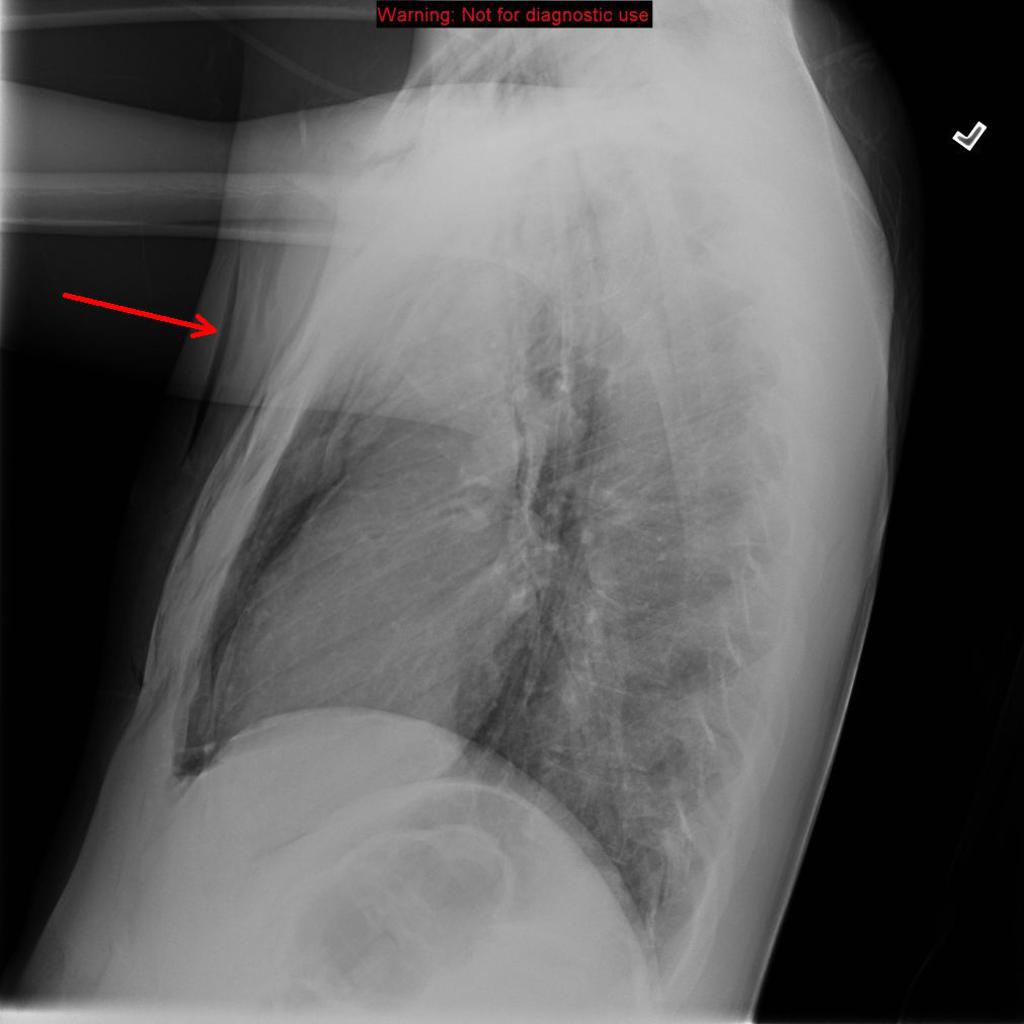
In spontaneous esophageal rupture (Boerhaave's syndrome), the diagnostic radiological finding is the V sign of Naclerio ( Figure 1 ), identified on a chest X-ray as two hypertransparent V-shaped lines, one along the left border of the aorta and the other creating the continuous diaphragm sign on the left.
Boerhaave Syndrome Radiology Key
Boerhaave's syndrome is the spontaneous rupture of the esophagus, which requires early diagnosis and treatment. Symptoms may vary, and diagnosis can be challenging. Case presentation Case 1: A 54-year-old Chinese man presented to us with sudden-onset epigastric pain radiating to the back following hematemesis.

Boerhaave syndrome Radiology Case Radiology
Boerhaave syndrome is a barogenic injury resulting from a sudden increase in intraluminal pressure against a closed cricopharyngeus. Neuromuscular dysfunction results in a non-relaxed cricopharyngeus with a resultant rise in pressure.

SciELO Brasil Boerhaave’s syndrome the role of conventional chest
The aim was to define the diagnostic value of chest radiography, esophagography, and computed tomography (CT) in patients with Boerhaave's syndrome. CT findings in 14 patients (11 male, 3 female; mean age: 60 years; median age: 66 years; age range: 36-78 years) with spontaneous esophageal perforation were retrospectively reviewed and compared to those of esophagography (n=11) and chest.
Boerhaave syndrome chest x ray wikidoc
Spontaneous oesophageal rupture (Boerhaave's syndrome) is an uncommon but serious condition. A retrospective review was undertaken of the management of 34 patients (age range 17-85 years) presenting between 1991 and 2006. Contrast swallow was possible in 22 patients, confirming the diagnosis in 17.

Boerhaave syndrome Radiology Case
Boerhaave syndrome is the perforation of the oesophagus caused by forceful vomiting due to increased intra-oesophageal pressure combined with relatively negative intra-thoracic pressure. This syndrome is associated with Mackler's triad (vomiting, chest pain, and subcutaneous emphysema).
Boerhaave Syndrome Radiology Key
Boerhaave syndrome refers to an esophageal rupture secondary to forceful vomiting and retching. Epidemiology It tends to be more prevalent in males, with alcoholism a risk factor. The estimated incidence is ~ 1:6000. Clinical presentation

Boerhaave syndrome Radiology Case
Boerhaave's syndrome is the spontaneous rupture of the esophagus, which requires early diagnosis and treatment. Symptoms may vary, and diagnosis can be challenging. Case 1: A 54-year-old Chinese man presented to us with sudden-onset epigastric pain radiating to the back following hematemesis. Upper gastrointestinal endoscopy revealed a full-thickness rupture of the esophageal wall.

Boerhaave syndrome Radiology Case
The aim was to define the diagnostic value of chest radiography, esophagography, and computed tomography (CT) in patients with Boerhaave's syndrome.

Boerhaave syndrome Radiology Case
Boerhaave's syndrome is a spontaneous rupture of the esophagus that occurs during intense straining. It most typically occurs during an episode of forceful or repeated vomiting. When the esophagus tears, toxic contents can leak out and cause infection. This is an emergency. Without treatment, it can be fatal within days.

Boerhaave syndrome Radiology Case
Boerhaave Syndrome: To Treat or Not to Treat by Means of Insertion of a Metallic Stent. Journal of Vascular and Interventional Radiology 6:5, 741-743. [Crossref] DC Cameron, J Black. 1995. Lightweight suction drainage - feeding system for oesophagogastric anastomotic leaks. Australasian Radiology 39:3, 314-319. [Crossref]
Boerhaave Syndrome Radiology Key
Boerhaave's syndrome is a rare form of esophageal perforation with a potentially complex presentation mimicking a variety of disorders. The prompt radiologic evaluation utilizing plain films, computed tomography and fluoroscopic esophagrams may avert the serious complications associated with a delay in diagnosis and treatment.

Boerhaave syndrome Radiology Case Git, Radiology
Emergency Radiology; Expert Panel Narrative Reviews; Global Reading Room; Journal Club; Noninterpretive Skills; Photon-Counting Detector CT; Point/Counterpoint; Special Series Review; Information. About AJR; Editorial Board;. Boerhaave syndrome: interventional radiologic management.
Boerhaave syndrome chest x ray wikidoc
Boerhaave syndrome represents the clinical syndrome associated with spontaneous esophageal rupture from retching and vomiting. The factor most often associated with a high mortality is a delay in diagnosis as this can result in significant mediastinal infection and tissue destruction [1-3]. Surgical repair remains the mainstay of therapy.